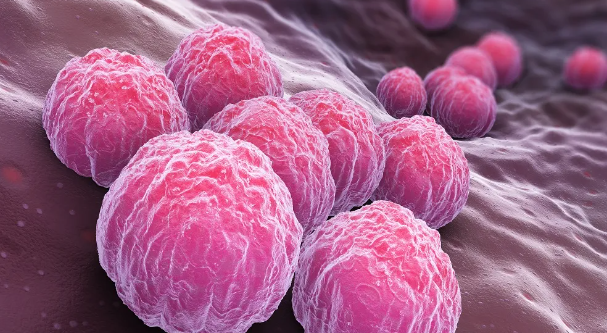
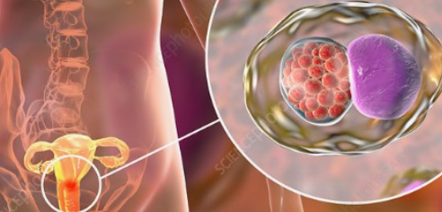
The initial and commonest form of cutaneous T cell lymphoma (CTCL) is mycosis fungoides [see clinical picture], which is characterized by the accumulation and clonal proliferation of malignant, epidermotropic, CD4+/CD45RO+ (helper/memory) T lymphocytes which interact with keratinocytes within the lesion. These keratinocytes are atypical in that they express ICAM-1 cell adhesion molecule plus class II major histocompatability complex. They also produce increased amounts of interleukin 1 (IL-1), which upregulates ICAM-1 expression on capillary endothelia, increases vascular permeability and enhances the effectiveness of other kertinocyte attractants for lymphocytes, such as IL-8. Thus these lesional keratinocytes have an enhanced ability to interact with epidermotropic, malignant T lymphocytes, which tend to produce a T-helper-2 cell cytokine profile. Loss of CD8+ cytotoxic T lymphocytes from the mycosis fungoides lesions is associated with poor disease progression and a poor prognosis (Ni et al., 2001).
Sezary syndrome is the leukaemic variant of CTCL, characterized by the presence of malignant monunclear cells in the blood. Sezary syndrome occurs most commonly in males. Clinically, the skin is diffusely bright red with scaling. There may be fever, chills, loss of weight and malaise. This may be accompanied by scaling and fissuring of the palms and soles, itching, baldness, ankle swelling and alterations to the nails (dystrophy). Histologically there is a perivascular accumulation of large number of atypical mononuclear cells. Diagnosis is made by skin biopsy and the finding of abnormal lymphocytes in the blood. Southern blot analysis of T-cell receptor gene rearrangements in CTCL in lesions often reveals the presence of a single neoplastic T cell clone, particlarly in later stages of the disease. Chemotherapy with nitrogen mustard may aid control of the disease, but is seldom curative.
Possible role of C. pneumoniae and other infections
It has long been suspected that CTCL might be caused by chronic local antigenic stimulation (Tan et al., 1974) with infection, including Staphylococcus aureus (Jackow et al., 1997) as well as C. pneumoniae (Abrams et al., 1999; 2001) a possible if controversial cause. Such localized bacterial infection might be expected to lead to the local production of inflammatory cytokines including interferon gamma (critical in immunity to, and immunopathology of, chlamydial infection) plus IP-10, a cytokine chemoattractive for epidermotrophic T lymphocytes (Sarris et al., 1995). Studies on the the growth requirements of the abnormal T lymphocytes in Sezary syndrome lead to the identification of a so-called Sezary cell activation factor (SAF) that stimulates the growth of both malignant and non-malignant T cells from this condition. Subsequent data suggested that SAF is a C. pneumoniae protein of approximately 30 kilodaltons, superficially at least resembling the C. pneumoniae T cell activation factor described by Halme et al., 1997. Skin biopsy specimens from patients with CTCL reacted similarly with monoclonal antibody to SAF, to the C. pneumoniae major outer membrane protein and to chlamydial lipopolysaccharide (see colour pictures in the full article version of Abrams et al., 1999). The expression of these antigens was reduced concommitant with clinical improvement in response to psoralen - long wavelength ultraviolet treatment of mycosis fungoides. Furthermore, C. pneumoniae genomic DNA, plus associated RNA transcription, was also detected in many of these antigen positive skin biopsies and C. pneumoniae was found able to replicate in keratinocytes (Abrams et al., 1999; 2001).
[Comment: A large number of theories abound concerning the cause of CTCL, which may well have several causes. However, these intriguing results suggest that at least one type of SAF may be a C. pneumoniae protein and indicate the association of C. pneumoniae antigens in the skin with CTLC. It cannot be inferred from these data that C. pneumoniae causes CTLC; here we are up against the same difficulty of distinguishing cause and effect that plagues analysis of the role of chlamydiae in other chronic diseases. One possibility, for example, is that C. pneumoniae antigen by chance is carried in circulating mononuclear cells to the skin lesions as a consequence of the chemoattraction of T lymphocytes to abnormal keratinocytes. It is unclear how chlamydiae might drive the clonal expansion of a selected T cell population. Despite these necessary caveats, this is interesting and innovative work which warrants further research because of the dual insights it may offer into CTCL and the pathogenesis of chronic chlamydial disease].
[MEW] March 2002
Abrams, J. T., Balin, B. J., Vonderheid, E. C. (2001). Association between Sezary T cell-activating factor, Chlamydia pneumoniae, and cutaneous T cell lymphoma. Annals of the New York Academy of Sciences 941, 69 - 85.
Abrams, J. T., Vonderheid, E. C., Kolbe, S., Appelt, D. M., Arking, E. J. & Balin, B. J. (1999). Sezary T-cell activating factor is a Chlamydia pneumoniae-associated protein. Clinical and diagnostic laboratory immunology 6, 895 - 905. Full article
Diamandidou, P., Cohen, P. R., Kurzrock, F. (1996). Mycosis fungoides and Sezary syndrome. Blood 88, 2385 - 2409.
Halme, S., Saikku, P. Surcel, H. M. (1997). Characterization of Chlamydia pneumoniae antigens using human T cell lines. Scandinavian Journal of Immunology 45, 378 - 384
Jackow, C. M., Cather, J. C., Hearne, V., Asano, A. T., Musser, J. M. & Duvic, M. (1997). Association of erythrodermic cutaneous T-cell lymphoma, superantigen-positive Staphylococcus aureus, and oligoclonal T-cell receptor V beta gene expansion. Blood 89, 32 - 40.
Ni, X., Parul, H., Zhang, C., Talpur, R. & Duvic, M. (2001). Fas ligand expression by neoplastic T lymphocytes mediates elimination of CD8+ cytotoxic T lymphocytes in mycosis fungoides: a potential mechanism of tumor immune escape? Clinical Cancer Research 7, 2682 - 2692.
Sarris, A. H., Esgleyes-Ribot, T., Crow, M., Broxmeyer, H. E., Karasavvas, N., Pugh, W., Grossman, D., Deisseroth, A. & Duvic, M. (1995). Cytokine loops involving interferon-gamma and IP-10, a cytokine chemotactic for CD4+ lymphocytes: an explanation for the epidermotropism of cutaneous T-cell lymphoma? Blood 86, 651 - 658
Tan, R. S. H., Butterworth, C. M., Mclaughlin, H., Malka, S. and Samman, P. D. (1974). Mycosis fungoides: a disease of antigen persistence. British Journal of Dermatology 91, 607 - 616